
Radiology
Radiology Treatment in Kolkata
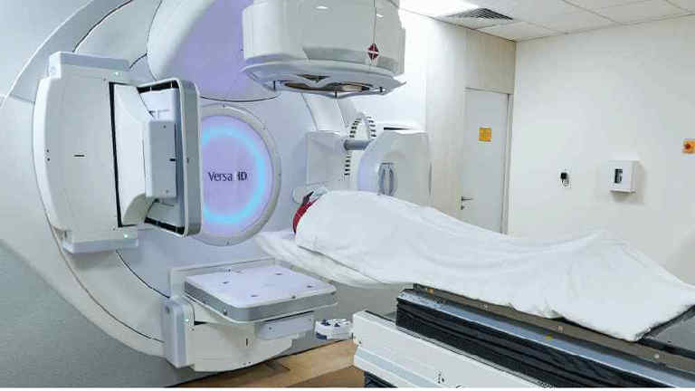
The Radiology Department at Manipal Hospitals in Broadway, Kolkata is equipped with cutting-edge equipment and has a highly qualified team of professionals who are committed to providing patients with accurate radiological diagnostics and comprehensive services. They improve patient care and treatment outcomes by ensuring comprehensive evaluations and rapid results with their advanced imaging equipment and experience. This dedication to quality highlights Manipal Hospitals standing as a preeminent medical facility offering first-rate radiology services.

OUR STORY
Know About Us
Why Manipal?
Manipal Hospitals stands at the forefront of modern radiology services, renowned for its commitment to superior medical care. Equipped with state-of-the-art technology, including MRI, CT scans, and X-rays, our advanced radiology services ensure precise diagnoses. Our team of highly skilled radiologists guarantees high-quality care by meticulously interpreting results, providing patients with accurate and timely insights into their health. With an extensive service offering, Manipal Hospitals can address a diverse range of medical needs under one roof, offering convenience and comprehensive care to our patients. From scheduling appointments to receiving results, we prioritise patient safety, comfort, and satisfaction at every step of the journey.
Through collaborative efforts with various specialisations, we tailor personalised and integrated treatment strategies to meet the unique needs of each patient. Our dedication to providing high-quality healthcare is evidenced by our numerous recognitions and accreditations, affirming our commitment to excellence.
Consult our radiology hospital if you need Radiology Treatment in Kolkata for treating disorders such as cancer. and other chronic conditions.
Treatment & Procedures
A frequent disorder called varicose veins is defined by twisted, bulging blood vessels that are visible beneath the skin's surface, usually in the legs, feet, and ankles. There may be pain and irritation associated with these veins. Varicose veins are sometimes accompanied by tiny red or purple streaks called spider veins. Severe occurrences of varicose…
The department of radiology provides imaging services for procedures such as stenting or angioplasty, wherein a catheter is guided into clogged arteries. A small balloon accompanies the catheter to open the artery, thereby allowing blood flow to occur.
Cancer is a global health concern, and various advancements and strategies are being implemented to improve cancer treatments as a top priority. A wide variety of therapeutic modalities are included in non-surgical cancer treatments, which are intended to fight cancer without requiring surgery. In the past two decades, the scope of non-surgical cancer…
A Biopsy is a procedure that involves the removal of a piece of tissue to diagnose any abnormalities or diseases. Specialists may suggest a Biopsy if they find any anomalies during a physical examination or through results obtained from any imaging tests. A Biopsy is also considered a gold standard for the diagnosis of cancer. Depending on the size…
Peripheral artery disease (PAD) is the accumulation of plaques (fat and cholesterol) within the legs or arms, making it harder for the blood to carry oxygen to tissue in the affected area. An individual with PAD develops a characteristic symptom of pain in the leg that starts with walking or exercise and goes away with rest.
A computed tomography (CT) scan is an imaging examination used by medical professionals to identify injuries and illnesses. It generates finely detailed pictures of your soft tissues and bones using a sequence of X-rays and a computer. Being a noninvasive test, you can undergo this procedure in a hospital or an imaging facility.
When a blood clot forms in one of the body's deep veins, it is known as deep vein thrombosis or DVT. This may occur if a vein sustains damage or blood flow is interrupted or ceases entirely. A lower-body injury and surgery involving the hips or legs are two of the most common risk factors for developing deep vein thrombosis (DVT).
Fluoroscopy is a medical imaging technique that displays internal organs and tissues in motion on a computer screen in real-time, using a series of rapid X-ray pulses. This technique records live images of the tissues within your body, providing continuous imaging during diagnostic and therapeutic procedures. Fluoroscopy is used by healthcare professionals…
A non-invasive medical imaging method called magnetic resonance imaging (MRI) creates precise pictures of the body's internal structures. Since MRIs do not use ionising radiation like CT scans or X-rays do, they are safer to use repeatedly. The patient lies down within a large, cylindrical magnet while radio waves are pointed at the body during the…
The kidney possesses several functions, including removing waste products and drugs from the body and maintaining hormone and body fluid levels. A disruption in these kidney functions could lead to the accumulation of toxins, which eventually leads to kidney failure. Although kidney failures can be addressed with Medications, specialists may recommend…
In some cases, regular radiology exams will not be able to obtain a specific image of the patient’s internal body and so nuclear medicine exams come into play. These types of exams use gamma rays along with a special camera to obtain the required images.
The sudden blockage of major arteries in the lung is caused by blood clots. The lungs can be damaged by these clots. Interventional radiology provides treatment in the form of catheter-directed thrombectomy/thrombolysis.
TIPS is a transjugular intrahepatic portosystemic shunt procedure th at is used to treat internal bleeding in the esophagus or stomach, for patients with cirrhosis. Imaging is used during the procedure to connect the portal and hepatic vein in the liver. A metal device known as a stent will hold the connection open, allowing blood flow from the bowel…
This therapy involves the use of cold, hot or chemical agents to treat cancerous tumors. Manipal hospital’s department of radiology specializes in the use of cryotherapy and microwave for tumor ablation.
A minimally invasive radiological technique called uterine fibroid embolisation (UFE) is used to treat symptomatic uterine fibroids, which are benign tumours in the uterus. Under UFE, which is performed by an interventional radiologist, a catheter is inserted into the uterine arteries via a tiny incision in the wrist or groin. The fibroids are then…
Radiographs, or X-rays, are among the basic imaging modalities used in radiology. They create two-dimensional pictures of the body's interior structure by using ionising radiation. Because X-ray imaging can be obtained quickly, is inexpensive, and is accessible, it is frequently used for diagnostic reasons. Electromagnetic radiation is sent through…

Our radiology department at Manipal Hospitals offers a wide range of services to accommodate various medical needs. We provide comprehensive diagnostics, from standard ultrasound tests to advanced DEXA scans. Our cutting-edge facilities provide accurate and precise fluoroscopy, nuclear medicine, and traditional X-ray services

Facilities & Services
Our radiology department at Manipal Hospitals offers a wide range of services to accommodate various medical needs. We provide comprehensive diagnostics, from standard ultrasound tests to advanced DEXA scans. Our cutting-edge facilities provide accurate and precise fluoroscopy, nuclear medicine, and traditional X-ray services. To further improve our diagnostic abilities, we specialise in echocardiograms, OB/GYN scans, and restricted vascular imaging.
Furthermore, we are proficient in using modern imaging methods to guide percutaneous abscess drainage treatments, biopsies, and mammography. We also excel in paracentesis treatments, making the most of our cutting-edge imaging modalities to provide the best possible care and results for our patients.
FAQ's
An extensive evaluation of the patient's health is done by a professional before any treatment or diagnostic treatments. This entails obtaining a thorough medical history from the patient and performing a thorough physical examination.
The specialist creates individualised treatment programs or suggests further diagnostic tests based on these findings. To maximise patient outcomes, our methodical approach provides precise diagnoses and customised treatment plans.
At Manipal Hospitals, prompt radiological scanning is essential for detecting a range of urgent medical issues. These include locating lumps, clots, and tumours in vital bodily parts like the brain, belly, and pelvis.
For early intervention and treatment planning, radiological imaging is also necessary for the detection of disorders such as internal bleeding within organs and liver cirrhosis. Our modern radiographic facilities allow for quick and precise diagnosis, prompt care of these serious illnesses, and better patient outcomes.
A variety of specialist tools are used at our radiological facilities to enable precise diagnostic imaging. Modern X-ray tubes are one example of this; they produce the radiation required for imaging operations. During examinations, radiographic tables give patients a stable and customisable surface, guaranteeing ideal placement for accurate imaging. Real-time images taken during procedures are displayed on monitors, enabling radiologists to evaluate image quality and make quick corrections.
Through the use of image brightness and contrast enhancers, fluoroscopy and other dynamic imaging studies can better visualise anatomical structures.
The majority of radiology exams are minimally invasive and created to be painless for the comfort of the patient. However, appropriate steps are taken to promote patient comfort when discomfort or pain is anticipated. A local anaesthetic may be used to numb the area being examined, or in certain cases, general anaesthesia may be used to produce temporary unconsciousness to manage discomfort, depending on the procedure and the patient's preferences. These methods are customised to meet the demands of each patient, ensure a pleasant and relaxing experience during radiological tests.
During the procedure, the patient may be positioned on a radiographic table or other specialised equipment to ensure optimal imaging. The technologist operates the imaging equipment, which may include X-ray machines, CT scanners, MRI scanners, or ultrasound machines, depending on the type of imaging needed. The imaging equipment captures detailed images of the internal structures of the body, which are then interpreted by radiologists. They analyse the images to make a diagnosis or provide information to the referring physician.
After interpretation, the radiologist generates a report detailing their findings, which is shared with the referring healthcare provider. Based on the results of the imaging study, further treatment or additional imaging studies may be recommended.
Test results from radiology are typically accurate when performed and interpreted by trained professionals using modern equipment. Radiological imaging techniques such as X-rays, CT scans, MRI scans, and ultrasound are highly sensitive and can provide detailed images of the body's internal structures.
However, occasional discrepancies may occur due to factors like patient movement or variations in interpretation. Overall, radiological tests are valuable diagnostic tools, but additional clinical information may be considered for comprehensive patient care.
While non-invasive imaging methods like ultrasounds and X-rays usually don't hurt, some treatments, including biopsies, can hurt a little because of pressure sensations and local anaesthetic. Despite being minimally invasive, injections guided by imaging can cause some mild discomfort at the injection site. Nonetheless, medical personnel put the comfort of their patients first and use strategies to reduce any pain they may feel while undergoing these treatments. Furthermore, patients are frequently informed in advance about the symptoms they can expect, so they are well-prepared.
Frequent yearly physical examinations are crucial for preserving general health and delaying the development of severe medical disorders later on. We support proactive health management and place a high priority on preventative treatment.
We advise patients to arrange yearly complete physical examinations to identify any possible health problems early and begin treatment as soon as possible. People can improve their long-term health and quality of life by putting prevention above cure. Our top goal is your health, and we are here to help you on your path to well-being.


