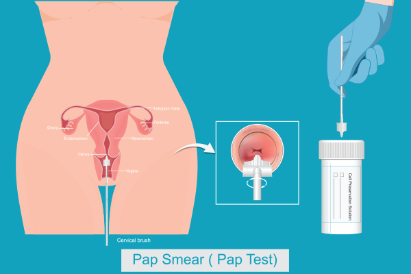
Ovarian cancer is one of the most challenging gynecological malignancies due to its often late diagnosis and complex treatment approach. The management of ovarian cancer varies significantly depending on whether the tumor is detected at an early stage (I-II) or in an advanced stage (III-IV). Understanding these differences is crucial for improving outcomes and ensuring patients receive the most effective treatment.
Synopsis
Early-Stage Ovarian Cancer (Stages I-II)
In early-stage ovarian cancer, the malignancy is confined to the ovaries or has limited local spread. The primary treatment strategy revolves around surgical removal of the tumour, often followed by chemotherapy to prevent a recurrence.
Surgical Treatment
Surgery is the cornerstone of early-stage ovarian cancer treatment. The goal is to remove all cancerous tissue while preserving as much normal function as possible. Common procedures include:
-
Total Hysterectomy: Removal of the uterus.
-
Bilateral Salpingo-Oophorectomy (BSO): Removal of both ovaries and fallopian tubes.
-
Omentectomy: Removal of the fatty tissue covering the abdominal organs.
-
Lymph Node Sampling: To check for possible cancer spread.
-
Fertility-Sparing Surgery: In select cases where cancer is confined to one ovary, a unilateral salpingo-oophorectomy (removal of one ovary and its fallopian tube) may be an option for women who wish to conceive in the future.
Chemotherapy
While surgery alone may be sufficient for some patients, those with high-risk features may require adjuvant chemotherapy. Platinum-based chemotherapy, usually a combination of carboplatin and paclitaxel, is the standard treatment to eliminate any remaining cancer cells and reduce recurrence risk.

Advanced-Stage Ovarian Cancer (Stages III-IV)
Advanced ovarian cancer signifies the spread of the malignancy beyond the ovaries, affecting the abdominal cavity or distant organs. Treatment requires a more aggressive, multi-modal approach.
Surgical Debulking (Cytoreduction)
The primary objective in advanced ovarian cancer surgery is cytoreduction, meaning the removal of as much tumour mass as possible. This is a crucial step, as optimal debulking (leaving no tumours larger than 1 cm) has been linked to better survival rates.
If the tumour is too extensive for immediate surgery, neoadjuvant chemotherapy (administered before surgery) may help shrink the tumour, making surgical removal more effective.
Chemotherapy and Targeted Therapy
-
Neoadjuvant Chemotherapy: Used when surgery is not initially possible.
-
Adjuvant Chemotherapy: Given after surgery to eliminate residual cancer cells.
-
Intraperitoneal Chemotherapy: Sometimes used to deliver chemotherapy directly into the abdomen for better-localized effect.
-
Targeted Therapy: Bevacizumab (an anti-angiogenesis drug) and PARP inhibitors are increasingly used, particularly in patients with BRCA mutations.
Comparison of Treatment Strategies
|
Treatment Modality |
Early-Stage (I-II) |
Advanced-Stage (III-IV) |
|
Surgery |
Hysterectomy, BSO, omentectomy, fertility-sparing options |
Cytoreductive surgery (debulking) |
|
Chemotherapy |
Adjuvant platinum-based therapy |
Neoadjuvant and/or adjuvant chemotherapy |
|
Radiation Therapy |
Rarely used |
Limited role, used for symptom relief |
|
Targeted Therapy |
Not typically required |
Bevacizumab, PARP inhibitors in selected cases |
Diagnostic Methods and Screening for Ovarian Cancer
Early detection of ovarian cancer remains a challenge due to its vague symptoms and the lack of routine screening tests. However, several diagnostic methods can aid in the identification and evaluation of ovarian cancer.
Imaging Tests
-
Ultrasound (Transvaginal and Abdominal): A non-invasive method to detect ovarian abnormalities, cysts, or tumours. Transvaginal ultrasound provides detailed images of the ovaries and surrounding structures.
-
CT Scan (Computed Tomography): Helps in assessing tumor size, spread, and involvement of nearby organs.
-
MRI (Magnetic Resonance Imaging): Offers a more detailed soft-tissue contrast than CT scans and is useful in distinguishing benign from malignant ovarian masses.
Blood Tests
-
CA-125 Marker: A commonly used tumour marker in ovarian cancer diagnosis. Elevated CA-125 levels may indicate ovarian cancer but can also be associated with benign conditions like endometriosis or pelvic inflammatory disease.
-
HE4 (Human Epididymis Protein 4): A more specific marker that, when combined with CA-125, improves the accuracy of ovarian cancer detection.
-
Other Biomarkers: Additional markers like CEA and AFP may be used in specific subtypes of ovarian cancer.
Role of Biopsy and Laparoscopic Evaluation
-
Biopsy: A tissue sample taken from the ovarian mass is examined under a microscope to confirm malignancy. In advanced cases, a biopsy may be taken from metastatic sites.
-
Laparoscopy: A minimally invasive surgical procedure that allows direct visualization of ovarian tumours and enables biopsy sampling for definitive diagnosis.
Conclusion
Manipal Hospital Gurugram is a leading healthcare institution offering state-of-the-art diagnostic and treatment facilities for ovarian cancer. With a multidisciplinary team of experts in surgical oncology, medical oncology, and radiation oncology, the hospital ensures personalized care tailored to each patient's needs.
Patients at Manipal Hospital Gurugram benefit from advanced technology, minimally invasive surgical options, targeted therapy, and holistic support services, making it a premier destination for ovarian cancer treatment in India.
Early detection and timely intervention are key to improving survival rates in ovarian cancer. If you or a loved one are experiencing symptoms or have concerns about ovarian cancer, consult with the specialists at Manipal Hospital Gurugram for expert guidance and comprehensive care.
FAQ's
Common symptoms include bloating, pelvic pain, loss of appetite, and frequent urination. These symptoms are often subtle and can mimic other conditions.
Diagnosis involves a pelvic exam, imaging tests (ultrasound, CT scan), blood tests (CA-125), and biopsy if necessary.
Early detection is difficult due to vague symptoms. Regular gynaecological check-ups and genetic testing for high-risk individuals can help.
Risk factors include a family history of ovarian or breast cancer, genetic mutations (BRCA1/BRCA2), age over 50, and never having been pregnant.
Preventive measures include genetic counselling, oral contraceptive use, and prophylactic removal of ovaries in high-risk individuals.


 5 Min Read


